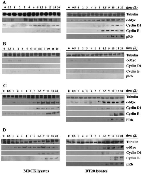
FIG. 5.
Induction of cell cycle proteins by continuous EGF exposure or by two pulses of standard and endosomal EGFR signaling. Subconfluent cultures of MDCK and BT20 cells were serum starved for 48 h and treated as indicated below. For each EGF treatment, cells were collected at the indicated times, and equal amounts of cell lysates were subjected to immunoblot analysis using antibodies to c-Myc, cyclin D1, cyclin E, and pRb. Tubulin antibody was used to assess protein loading. (A) Cells were treated continuously with EGF (100 ng/ml) until assayed. (B) Cells were treated for 1 h with EGF, after which unbound ligand was removed and cells were cultured in starvation medium. (C) Cells were treated with two 1-h pulses of EGF administered at 0 and 8 h, and pulses were terminated by washing as described above. (D) Cells were treated with AG1478 and EGF and then acid stripped of recycled ligand and washed free of AG1478 to activate internalized EGF-EGFR complexes. At 8 h, the same treatment was repeated. MDCK lysates were not analyzed for pRb phosphorylation, because the antibody didn't detect the canine protein.

