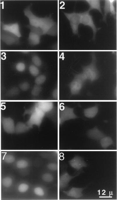
Figure 1.
Smad 3 MH1 domain is constitutively localized to the nucleus after both transient and stable expression. (1–6) Transient expression; MH1 and MH2 domains of Smad 3 and Smad 4 proteins were fused to the C terminus of eGFP and transiently expressed in BOSC cells. One day after transfection, the living cells were photographed under the fluorescence microscope to detect the GFP signal. (1) GFP; (2) GFP-Smad 3 full-length protein fusion; (3) GFP-Smad 3 MH1; (4) GFP-Smad 3 MH2; (5) GFP-Smad 4 MH1; and (6) GFP-Smad 4 MH2. Nuclear localizations were confirmed by 4′,6-diamidino-2-phenylindole staining of fixed cells (not shown). (7–8) Stable expression. The MH1 domains of Smads 3 and 4 each were fused with GFP and cloned into the retroviral pMX vector. Two days after transfection into BOSC cells, virus-containing media were collected and used to infect L20 cells. Two days later, living cells were photographed under the fluorescence microscope. (7) Smad 3 MH1; (8) Smad 4 MH1.

