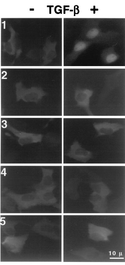
Figure 4.
NLS-mutated Smad 3 proteins failed to translocate to nucleus in response to TGF-β. L20 (Mv1Lu) cells stably expressing wild-type or mutant forms of GFP-Smad 3 were first starved in low-serum medium (0.5% FCS in DME) for 2–3 h and then treated with 200–500 pM TGF-β. After 1 h, the cells were then photographed under the fluorescence microscope. Left for each sample shows the image before stimulation; Right shows the image after TGF-β addition. (1) Wild-type Smad 3; (2) Smad 3 K43N/K44Q; (3) Smad 3 ΔK43K44; (4) Smad 3 K44E; (5) Smad 3 ΔK40K41.

