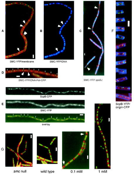
FIG. 1.
Fluorescence microscopy of B. subtilis cells growing at mid-exponential phase. (A to D) SMC-YFP (green), membranes (red; A and C), DNA (blue; B and C), and tau subunit of DNA polymerase (red; D). (A and B) Strain JM24 (smc-yfp); arrowheads indicate SMC at midcell or in a bipolar location. (C) Strain JM28 (smc-yfp spo0J::spec); arrowheads indicate the absence of SMC fluorescence in anucleate cells, which arise at a frequency of about 1% in spo0J mutant cells. (D) Strain JM27 (smc-yfp dnaX-cfp); arrowheads indicate SMC foci flanking a central DNA polymerase focus. (E) Strain PG44 (scpB-cfp smc-yfp); upper panel shows CFP fluorescence, middle panel shows YFP fluorescence, and lower panel shows an overlay. (F) Representative cells showing the localization of ScpB-YFP (red), origin regions (green), and membranes (blue). (G) DNA (green) and membranes (red); first panel shows strain PGΔ388 (smc::kan), second panel shows strain PY79 (wild type), third panel shows strain CAS5 (Phyperspac-smc) with 0.1 mM IPTG, and fourth panel shows strain CAS5 (Phyperspac-smc) with 1 mM IPTG. Arrowheads in the third panel indicate increased DNA-free spaces in cells. Ends of cells are indicated by thin white lines. Thick white bars, 2 μm.

