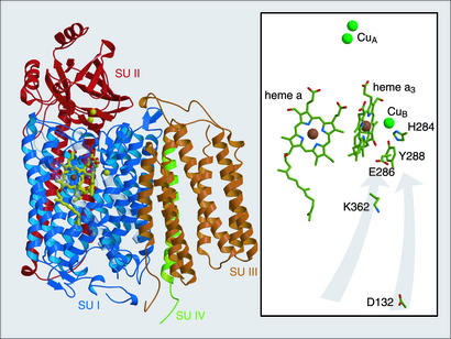
Fig. 1.
Structural view of CcO from R. sphaeroides (PDB ID 1M56; ref. 9) parallel to the membrane surface, with periplasmic surface on top. (Left) Ribbon diagram highlighting the arrangement of the four subunits (SU I–IV). (Right) A view in which the protein backbone is stripped to show the chemical structure of particularly important catalytically active groups. The gray arrows indicate the two spatially separated proton transfer pathways that contain the critical residues D132 and K362.

