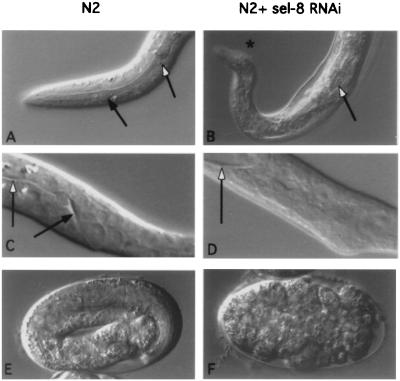
Figure 3.
RNA-mediated interference. Nomarski photomicrographs of the progeny of untreated or mock-injected N2 hermaphrodites (Left) and the progeny of hermaphrodites that had been injected with sel-8 dsRNA (Right). (A and B) Head region, L1 larvae. Black arrow indicates the anterior bulb of the pharynx (missing in B), and the white arrow indicates the posterior bulb. (C and D) Tail region, L1 larvae. Black arrow indicates the rectum (missing in D), and the white arrow indicates the intestine. Note the distension of the intestine in D resulting from the absence of the rectum. (E and F) Embryos 6–7 h after egg laying. (E) The embryo has reached the 3-fold stage. (F) The embryo has arrested without undergoing elongation. We also observed embryos with the same overall morphology as in F when we injected N2 hermaphrodites with a mixture of glp-1 and lin-12 dsRNAs, or with lag-1 dsRNA.

