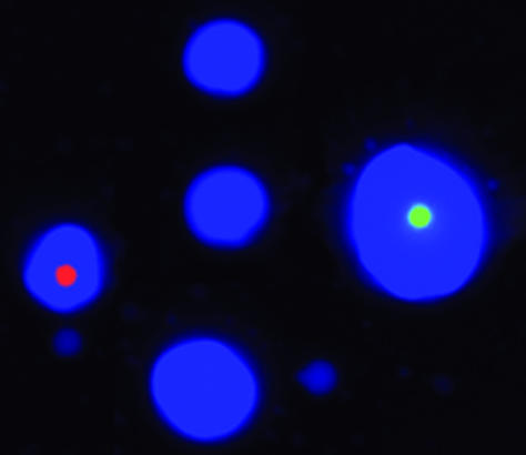
Fig. 2.
Photograph of a typical microemulsion. Microemulsions were made as described in Materials and Methods with the exception that the aqueous compartments contained cascade blue-labeled dCTP and the beads were prelabeled by binding to oligonucleotides coupled to R-phycoerythrin (red) or Alexa 488 (green). One microliter of microemulsion was deposited in 1 μlofoil on a microscope slide before photography. Of the seven aqueous compartments visible in this picture, two contain beads. Note the heterogeneous size of the aqueous compartments (beads are 1.05 μm in diameter).

