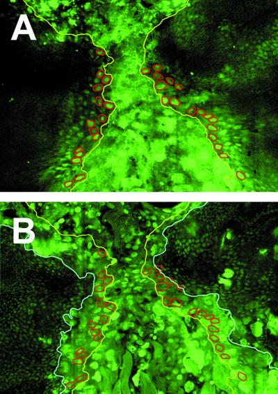
Figure 2.
Imaginal cells spread over larval cells during thorax closure. Confocal images of a 5-h APF pupa expressing a nuclear GFP (GFPn) under the control of Arm-Gal4. GFPn is expressed in all imaginal (small diploid nuclei) and larval (large polyploid nuclei) cells. (A) Dorsal surface focal plane. Leading-edge imaginal cells are highlighted in red. The edge of the spreading discs is marked in yellow. (B) A focal plane situated 6 μm below the dorsal surface. The edge of the underlying larval epidermis is marked in light blue.

