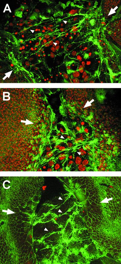
Figure 3.
Imaginal cells extend filopodia that connect contralateral discs. (A) Confocal image projection of a 5-h APF pupa stained with phalloidin to label polymerized actin and TO-PRO3 to mark nuclei. Long, thick filopodia extend from the wing imaginal disc edges, expand over the larval tissue, and eventually connect the confronting discs (arrowheads). (B) At 6 h APF, after discs contacted anteriorly, filopodia (arrowheads) extend from more posterior areas of the leading edge (confocal projection). (C) Dorsal surface confocal image of a 6-h APF pupa. Filopodia link the contralateral discs, and imaginal cells change shape extending toward the midline.

