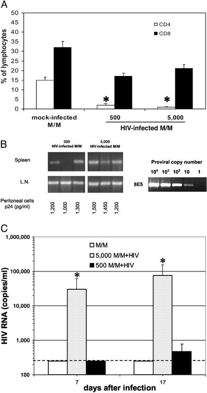
Fig. 2.
Few M/M are sufficient to efficiently spread HIV infection in hu-PBL-SCID mice. (A) Percentage of CD4+ T lymphocytes (open bars) and CD8+ T lymphocytes (filled bars) were measured in hu-PBL-SCID mice at day 21 after challenge with 500 or 5,000 HIV-infected macrophages, or 5,000 mock-infected macrophages; *, P < 0.01 compared with CD4 of mice challenged with uninfected M/M. (B) HIV-1 proviral copies were evaluated in spleen and lymph nodes (L.N.) by DNA PCR; as a control, several dilutions of DNA from 8E5 HIV-infected cells are reported. Cocultivation of peritoneal cells with donor autologous PBLs was also performed. (C) HIV-1 RNA was measured in the plasma of hu-PBL-SCID mice at days 7 and 17 after challenge with 5,000 HIV-infected macrophages (5,000 M/M+HIV, stippled bars), 500 HIV-infected macrophages (500 M/M+HIV, filled bars), or 5,000 mock-infected macrophages (M/M, open bars); *, P < 0.001 compared with CD4 of mice challenged with uninfected M/M. The dashed line defines the threshold of HIV-RNA detection. Three mice were used in each group.

