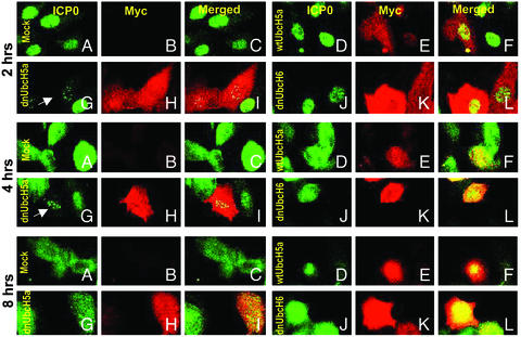
Fig. 5.
Distribution of ICP0 in SK-N-SH cells exposed to insect cell medium or baculoviruses encoding wtUbcH5a, dnUbcH5a, or dnUbcH6 and infected with HSV-1(F). The procedure for the treatment of the slide cultures was the same as described in Fig. 4 except that the cells were fixed at 2, 4, or 8 h after HSV-1(F) infection and reacted with anti-ICP0 (FITC) and anti-myc (Texas red) antibodies. The arrows point to cells containing ICP0 in speckled nuclear structures. In other assays these speckled structures were juxtaposed to or overlapped ND10 structures (data not shown).

