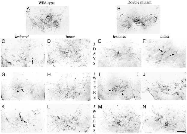
Fig. 3.
Immunochemical detection of serotonergic system below the lesion level. In intact wild-type (A) and double mutant (B) mice, 5HT-IR fibers are present all over the ventral horn of the cord and outline the motoneurons. Three days after hemisection (C–F), a substantial decrease of 5HT-IR was observed in the ventral horn on the lesioned side (C and E) compared with the intact side (D and F), in both wild-type (C and D) and double mutant (E and F) mice. This decreased 5HT-IR was more dramatic in both ventral horns of the double mutant mice (E and F, arrows). Three weeks after hemisection (G–J), a moderate increase of 5HT-IR with coarse fibers was observed in the ventral horn on the lesioned side in wild-type mice (G, arrow), whereas a substantial increase of 5HT-IR was observed on the lesioned side of double mutant mice with thin fibers delineating the motoneurons (I, arrowheads). 5HT-IR on the intact side of wild-type (H) and double mutant (J) mice tends to return progressively to control values. Five weeks after the lesion (K–N), scarce IR fibers were observed invading the ventral horn on the lesioned side in wild-type mice (K, arrows). In contrast, numerous thin 5HT-IR fibers coming from the intact side can be seen on the lesioned side of a double mutant mouse (M) where they outline their target, the motoneurons. 5HT-IR on the intact side went back to control value in both wild-type (L) and double mutant (N) mice and outlines their target, the motoneurons. (Bar = 75 μm.)

