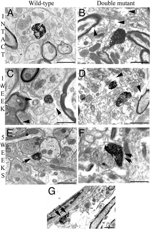
Fig. 5.
Ultrastructural organization and serotonergic profiles in the SC below the lesion level. Electron micrographs of a wild-type (A, C, and E) and double mutant (B, D, and F) mouse. An extensive extracellular space can be seen in an intact double mutant mouse (B, arrowheads) as compared with that of an intact wild-type animal (A). One week after the lesion (C and D), the ultrastructure on the lesioned side is altered in a wild-type mouse (C, arrowheads) but more so in the double mutant one (D, arrowheads). Five weeks after the lesion (E and F), few 5HT-IR profiles can be seen in a wild-type mouse (E, arrow), whereas in a double mutant mouse, more abundant 5HT-IR boutons make synaptic contact on dendrites (F, arrows) or eventually are directly apposed on the basal lamina of blood vessels (G, arrows). (Bars = 1 μm.)

