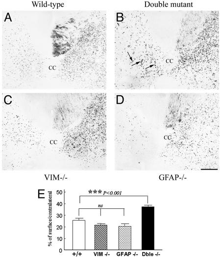
Fig. 6.
Anterograde labeling of CST by wheat germ agglutinin-conjugated horseradish peroxidase tracing. (A–D) Transverse sections of SC below the lesion level. Few labeled fibers were seen on the lesioned side of the SC in wild-type (A), Vim-/- (C), and GFAP-/- (D) mice. Observe in a double mutant mouse (arrows, B) a significant number of thin fibers that crossed the midline from the intact side to the lesioned side reinnervating the denervated gray matter. (Bar = 50 μm.) (E) Quantification of corticospinal sprouting. Fiber density on the lesioned side is expressed as a percentage of that of the intact side and illustrates a significant sprouting of corticospinal fibers only in double mutant mice. ***, P < 0.001, Mann–Whitney U test. Error bars indicate standard errors.

