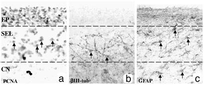
Fig. 3.
PCNA+, βIII-tubulin+, and GFAP+ cells are located in different regions within the SEL as demonstrated with bright-field immunohistochemical serial sections through the EP, SEL, and CN of a grade 2 HD brain. (a) PCNA+ cells are evenly distributed within the SEL (arrows demonstrate examples of PCNA+ cell bodies), and the EP is also densely stained. (b) βIII-tubulin+ fibers and cell bodies are present in the lower part of the SEL (arrows indicate cell bodies with immunoreactive fibers). (c) GFAP+ cells are distributed homogenously throughout the SEL and in the CN (arrows indicate examples of GFAP cell bodies).

