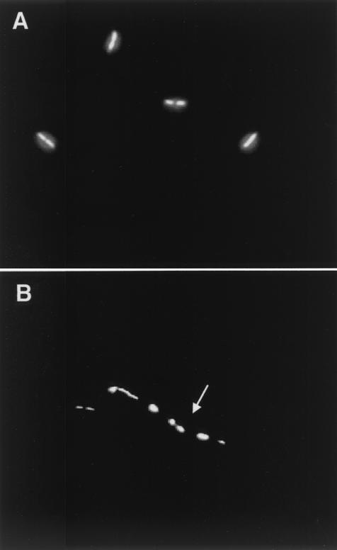
FIG. 2.
Nucleoid distribution in recA mutants of T. thermophilus. Shown is fluorescence microscopy of exponential cultures of the wild type (A) and the ΔrecA mutant (B) of T. thermophilus HB27. The cells were stained with DAPI. Note the irregular distribution of the nucleoids (arrow) in a filament of the mutant strain.

