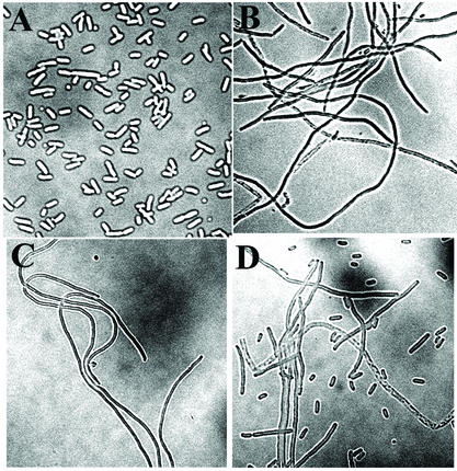
FIG. 4.
Effects of minE mutations on cell division patterns. Micrographs of Nomarski images of RC1 containing pSY1083 (Plac-minC minD minE) (A), pEM22S (Plac-minC minD minEL22S) (B), pEM25 (Plac-minC minD minEI25R) (C), and pEM19 (Plac-minC minD minEK19A) (D) are shown. The cells were prepared and analyzed as previously described (1).

