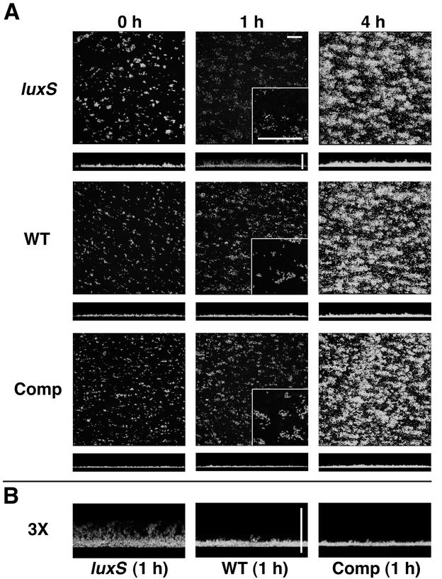
FIG. 7.
S. gordonii luxS mutant biofilm phenotype in saliva. (A) Time courses (left to right) of biofilm development in saliva-conditioned flow cells by luxS mutant (luxS), wild-type (WT), and complemented luxS mutant (Comp) S. gordonii strains. x-z reconstructions of each biofilm are shown below each x-y image. Digital zooms (magnification, 3×) of the lower left corner of each 1-h x-y image are shown as insets. (B) Digital zooms (magnification, 3×) of the center of each 1-h x-z reconstruction. Scale bars, 50 μm.

