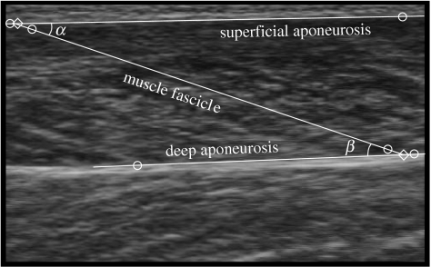
Figure 2.
Ultrasound image from the medial gastrocnemius during cycling at a pedal speed of 60 r.p.m. and a crank torque of 17 N m. The circles show the digitized points, and the lines show the interpolated aponeurosis and fascicle trajectories. The diamonds show the ends of a muscle fascicle that has a pennation angle of α with the superficial aponeurosis. The angle between the superficial and deep aponeuroses is β−α.

