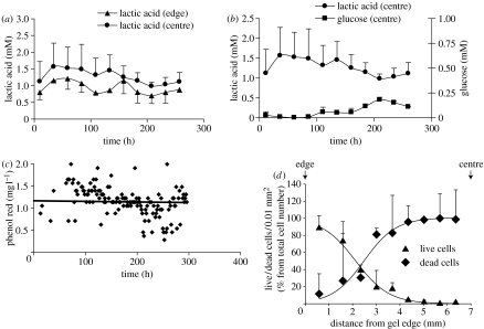
Figure 5.
Use of probes to detect cell death in the centre of a gel. (a) Cells were introduced into a gel, diameter 20 mm, thickness 13.5±0.1 mm, at an initial concentration of 15×106 cells cm−3. Solutes diffused from both sides of the gel to the gel centre. Two probes (‘edge’ probes) were introduced symmetrically, each at 3.2±0.1 mm from the nearest surface of the gel; two more probes (‘centre’ probes) also symmetrically, at 5.8±0.1 mm from the nearest surface of the gel. The medium supplied to the bioreactor contained initially 1 g l−1 glucose, 25 mM HEPES and its osmolarity was 380 mOsmol. The concentration of HEPES in the medium was subsequently increased to 50, 75 and 100 mM in order to maintain pH 7.4. Dialysate samples were collected from the probe every 99 min. The concentrations of 15 samples were averaged over 24.75 h periods. (b) Local glucose and lactic acid concentrations were determined by the probe in the centre of the bioreactor. Lactic acid local concentration correlated negatively with the level of glucose (r=−0.74). (c) Measurement of probe fouling by monitoring the Phenol Red concentration in the dialysate. Results show that the Phenol Red concentration in both probes remained constant over the period of the experiment (250 h). (d) The cell viability profile after two weeks of culturing in the bioreactor showing that viable cells were only seen within a 5 mm zone at the edges of the gel.

