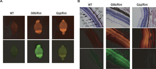
FIGURE 5.
ISS dependent exon IIIb silencing in neural tissues. (A) Fluorescent images of whole brains from wild-type (WT), GIIIb/Rint, and GΔΔ/Rint animals were obtained using a fluorescence light box and acquired for 300 msec with a CCD camera using RFP (upper panels) or GFP (lower panels) excitation/emission filters. The yellow signal observed in the GFP channel when imaging GΔΔ/Rint was due to bleed-through from RFP; it was not observed in GΔΔ/rosa26+ mice (not shown). (B) Cryosections of the eyes were stained with H&E and imaged by light microscopy to detect histological structures (top panels) after they were analyzed by epifluorescence microscopy to detect RFP (middle panels) and GFP expression (bottom panels). Eye sections illustrate a detailed view of the retina. All images in B were acquired at 200× magnification. (GC) layer of ganglion cells; (IPL) inner plexiform layer; (INL) inner nuclear layer; (OPL) outer plexiform layer; (ONL) outer nuclear layer; (R&C) rods and cone receptors; (PEp) pigment epithelium; (Ch) choroids.

