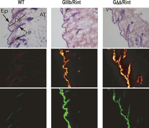
FIGURE 6.
ISS independent silencing in the skin. Cryosections of skin from the back of the animals were stained with H&E and imaged by light microscopy to detect histological structures (top panels) after they were analyzed by epifluorescence microscopy to detect RFP (middle panels) and GFP expression (bottom panels). Images were acquired at 200× magnification. (AT) adipose tissue; (De) dermis; (Ep) epidermis; (HF) hair follicle.

