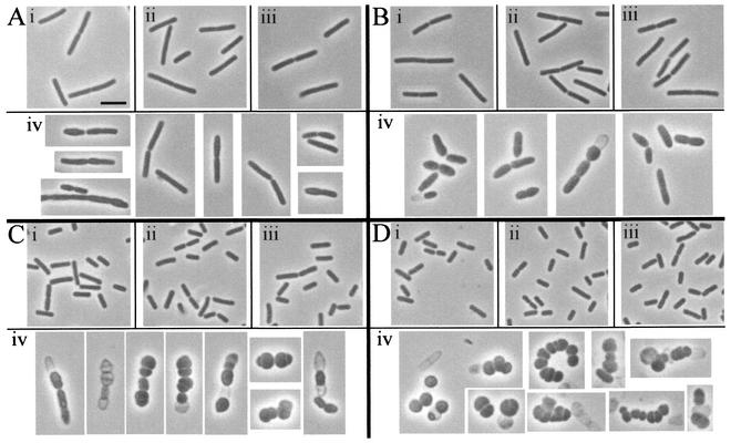
FIG. 5.
Phase contrast microscopy of vegetative cells. Cultures were grown in the presence of xylose and then resuspended in medium containing or lacking xylose, as described for Fig. 3. Culture samples were examined 80 min (A), 120 min (B), 180 min (C), and 240 min (D) after resuspension. Cells are from the wild-type strain PS832 with (i) and without (ii) xylose and the pbpA pbpH amyE::xylAp-pbpH strain DPVB207 with (iii) and without (iv) xylose. All images are at the same magnification. Scale bar in Ai, 2 μm.

