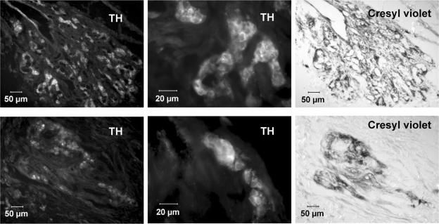
Figure 2. Immunostaining of central portion CB sections for TH with cresyl violet counterstaining.
Upper row, sections from a control CB. Lower row, sections from a hyperoxic CB. Both sections were immunostained for TH, the images recorded, then the sections were counterstained with cresyl violet to reveal the general histology, particularly the lumen of the capillaries. The middle sections correspond to magnified areas, demonstrating the intensity and localization of the immunostaining in type I cell cytoplasm.

