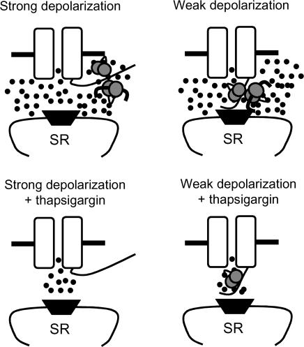
Figure 8. Schematic depiction of the hypothesized relationship between cell membrane potential, L-type Ca2+ channel C terminus motion and ‘sensing’ by CaM and CaMK.
The L-type Ca2+ channel pore forming subunit (α1C) is shown as a pair of open rectangles with a central pore. The C terminus protrudes into the cytoplasmic space from the right rectangle and the pore region and SR Ca2+ release channel (ryanodine receptor, dark trapezoid) are en face. Ca2+–CaM is depicted as a pair of stippled circles linked by a curved segment and activated CaMK is shown as a thick bar with curved ends bound to Ca2+–CaM. According to the hypothesized model, strong depolarizations motivate the C terminus to move away from the α1C pore so that sensing is primarily from the SR. Weak depolarizations leave the C terminus in the vicinity of the pore where sensed is directly from ICa−L. Inactivation of SR Ca2+ release (by thapsigargin) results in significant impairment of sensing through CaM and CaMK during strong depolarizations, while from ICa−L is sufficient for Ca2+–CaM at weak depolarizations in the absence of SR Ca2+ release.

