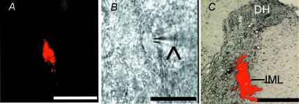
Figure 1. identification of retrogradely labeled spinally projecting PVN neurons.
A, a PVN neurone in the brain slice labelled with FluoSphere identified with fluorescence illumination. B, the same neurone recorded with a glass electrode (∧) viewed with Nomarski optics. C, photomicrograph showing the FluoSphere injection site (red) at the spinal intermediolateral cell column (IML) in one rat. Note that the bright field and fluorescence images were taken from the same tissue section and superimposed to show the location and diffusion of the FluoSphere injection. Scale bar, 50 μm in A and B; 500 μm in C. DH, dorsal horn.

