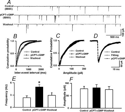
Figure 6. Lack of effect of pCPT-cGMP on mEPSCs in labeled PVN neurons.
A, raw tracings showing the spontaneous mIPSCs during control and application of 30 μm pCPT–cGMP in the presence of 100 μm IBMX in a labelled PVN neurone. B and C, cumulative plot analysis of mIPSCs of the same neurone as in A showing the distribution of the interevent interval (B) and peak amplitude (C) during control and pCPT–cGMP application in the presence of IBMX. pCPT–cGMP decreased the interevent interval of mIPSCs (Kolmgorov-Smirnov test, P < 0.05) without changing the distribution of the amplitude. D, superimposed averages of 100 consecutive mIPSCs obtained during control and pCPT–cGMP application. The decay time constants during control (τfast= 4.79 ms and τslow= 23.12 ms) and pCPT–cGMP administration (τfast= 4.74 ms and τslow= 22.25 ms) were similar. E,F, summary data showing the effect of pCPT–cGMP on the frequency (E) and amplitude (F) of mIPSCs of nine labelled PVN neurones. Data presented as means ±s.e.m.*P < 0.05 compared to the control (Kruskal-Wallis test).

