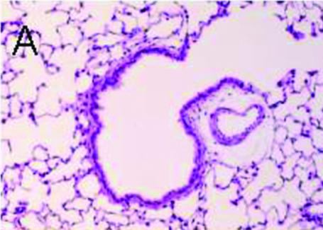
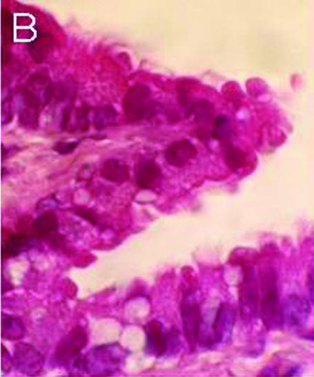
Figure 1. Histological examination of rat lung (HE staining).
A, low magnification. Terminal bronchiole and alveolar cavity. The terminal bronchiolar surface is lined with ciliary cells, but not the alveolar surface. B, high magnification. Ciliary cells with cilia on the apical surface line the luminal surface of the terminal bronchiole.


