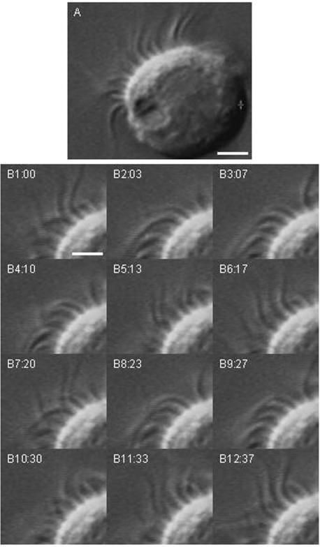
Figure 2. Video DIC images of a single ciliary cell.
A, the differential interference contrast (DIC) image shows beating cilia located on the apical surface of the cell. Each cilium movement was detected in the video image. Scale bar, 4 μm. B, frame images taken every 30–40 ms. Panels B1–B12 show two beating cycles (panels 1–6 and panels 7–11). The ciliary beat frequency of this cell is approximately 5.4 Hz. Scale bar, 2 μm.

