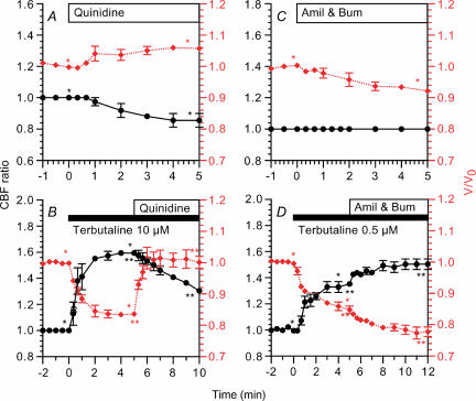
Figure 7. Changes in CBF and cell volume induced by inhibition of K+ channels (quinidine) or Na+ entry pathways (amiloride and bumetanide).
CBF ratios (•) are plotted on the left y-axis and V/V0 values (⋄ (red)) are on the right y-axis. A, quinidine (500 μm) decreased CBF ratio and increased V/V0 in unstimulated bronchiolar ciliary cells. B, in 10 μm terbutaline-stimulated cells, the subsequent addition of quinidine (500 μm) increased V/V0 immediately and decreased CBF gradually. C, amiloride (Amil) and 20 μm bumetanide (Bum) decreased V/V0 gradually, but did not change CBF in unstimulated cells. D, in 0.5 μm terbutaline-stimulated cells, the subsequent addition of both 1 μm amiloride and 20 μm bumetanide enhanced cell shrinkage and CBF increase. *,**Paired values significantly different (P < 0.05).

