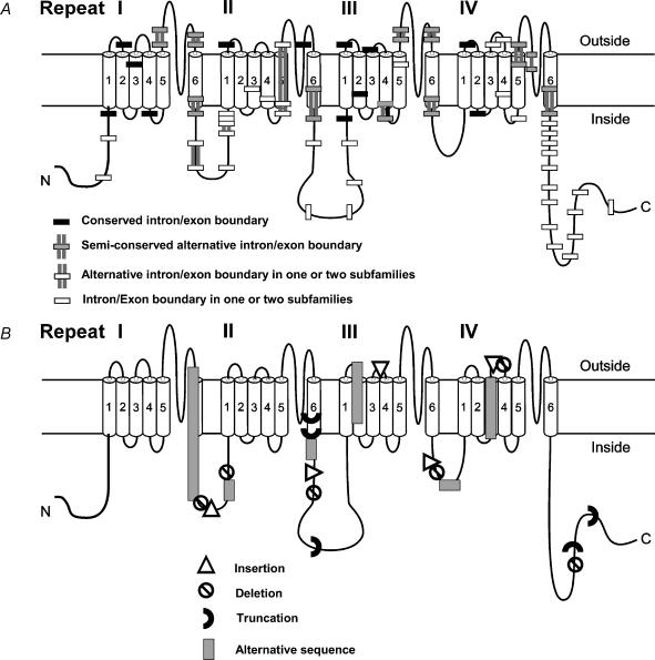
Figure 2. Scheme of the voltage-gated calcium channel α1 subunit.
The α1 subunit of voltage gated calcium channels consists of four domains (repeats) of six transmembrane segments connected by intracellular loops. A, conservation of the gene structure at the protein level as determined by protein alignments encoded by all exons of all 10 members of the CACNA1 family. Each protein region encoded by an exon is delineated by bars. White, black and grey bars indicate degree of conservation as noted in the figure. The following human reference sequences were used at NCBI: Cav2.1: NP_075461; Cav2.2: NP_000709; Cav1.2: NP_000710; Cav1.3: NP_000711; Cav2.3: NP_000712; Cav1.4: NP_005174; Cav3.1: NP_061496; Cav3.2: NP_066921; Cav3.3: NP_066919; and Cav1.1: NP_000060. B, regions of the protein that are affected by alternative splicing. Note that the changes now reflect the protein level only (i.e. deletions leading to frame shifts and early truncations are marked as truncation only). The diversity of the primary protein sequence due to insertions, deletions, truncations and alternative sequences is marked by the various symbols.

