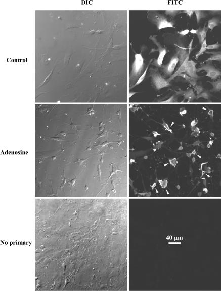
Figure 1. Fluorescence labelling of cultured rat pituicytes with antitaurine antibody.
Left panels are differential interference contrast pictures; right panels are the corresponding FITC pictures showing antitaurine labelling. Upper panels show a standard pituicyte culture (1 h in 0.025% serum) with mostly flat or elongated cells. Middle panels show another culture to which 10 μm adenosine was added to induce pituicyte stellation (Rosso et al. 2002a). Most cells have shrunken and roundish somas with numerous processes. Arrowheads point to cells in which taurine displays clear submembrane localization. Bottom panels show a mixed culture (with both flat and stellate pituicytes) in which primary but not secondary antibody was omitted. All pictures were taken with a 20 × objective lens.

