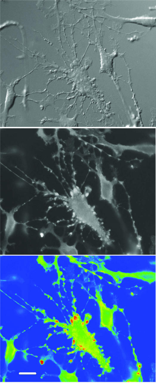
Figure 4. Selective concentration of taurine in pericellular protrusions.
Upper panel shows a differential interference contrast picture of stellate pituicytes 1 min after 10 nm VP application. The centre cell displays intense reorganization with formation of pericellular protrusions. This asynchronous process is not observed in all pituicytes simultaneously. Middle panel shows corresponding FITC fluorescence revealing taurine labelling. Lower panel shows pseudo-colour conversion of FITC fluorescence displaying low-to-high (blue-to-red) intensity. Pericellular protrusions selectively light up in red. Pictures were taken with a 40 × objective lens. Calibration = 16 μm.

