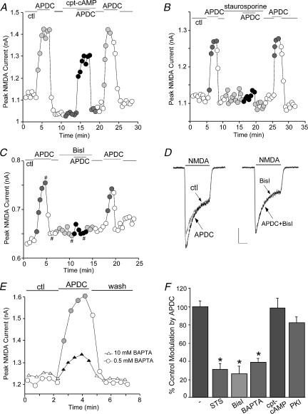
Figure 4. The mGluR2/3 enhancement of NMDAR currents was dependent on PKC, but not PKA.
A, B and C, plot of peak NMDAR currents as a function of time and drug application. Application of the broad-spectrum kinase inhibitor staurosporine (1 μm, B) or the specific PKC inhibitor bisindolylmaleimide (Bisl, 1 μm, C), but not the PKA activator cpt-cAMP (50 μm, A), largely blocked the APDC-induced enhancement of NMDAR currents. D, representative current traces taken from the records used to construct C (at time points denoted by #). Scale bars: 0.15 nA, 0.5 s. E, plot of peak NMDAR currents in neurones dialysed with internal solutions containing low (0.5 mm) or high (10 mm) concentrations of BAPTA. APDC had significantly smaller effect in the neurone loaded with high BAPTA. F, cumulative data (mean ±s.e.m.) showing the percentage control modulation of NMDAR currents by APDC in the absence (n= 12) or presence of staurosporine (STS, n= 16), bisindolylmaleimide (Bisl, n= 19), high BAPTA (n= 13), cpt-cAMP (n= 10) or the PKA inhibitor PKI[14–22] (0.1 μm, n= 9). *P < 0.005, anova.

