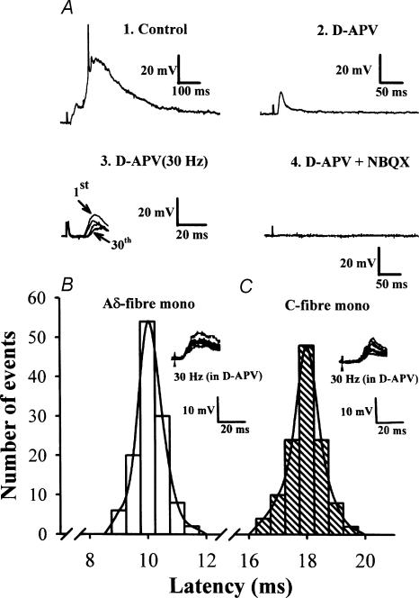
Figure 1. EPSPs recorded from trigeminal caudal neurones in response to mandibular nerve stimulation.
A, EPSPs evoked in the trigeminal caudal neurones by stimulating the mandibular nerve at 0.033 Hz (1, 2, and 4) and at 30 Hz (3) in the presence of strychnine sulphate (0.5 μm) and bicuculline methiodide (10 μm). Perfusing the slices with NMDA receptor antagonist d-APV (50 μm) largely attenuated slow EPSP components (2) and further addition of the non-NMDA receptor antagonist NBQX (20 μm) completely abolished the synaptic response (4). Note that the short-latency fast EPSP component recorded in the presence of d-APV (5 traces superimposed) persisted up to 30 Hz with a relatively stable latency with no failures (3). The resting membrane potential of this cell was –67 mV. B and C, monosynaptic nature of EPSPs remaining after high-frequency repetitive stimulation in the presence of d-APV (50 μm). The latency histograms of EPSPs evoked at 30 Hz in two different trigeminal neurones are shown. From the stable latency and absence of failure during 30 Hz stimulation, we deduced that these afferent inputs are monosynaptically connected. Inset in B, representative monosynaptic Aδ-fibre EPSPs (6 traces superimposed) in one neurone had a low threshold (4 V; 0.3 ms) and short latency (10.6 ms). Inset in C, representative monosynaptic C-fibre EPSPs (6 traces superimposed) in one neurone had a high threshold (11 V; 0.3 ms) and long latency (18.6 ms). The latencies (120 responses each in B and C) were fitted with a Gaussian distribution.

