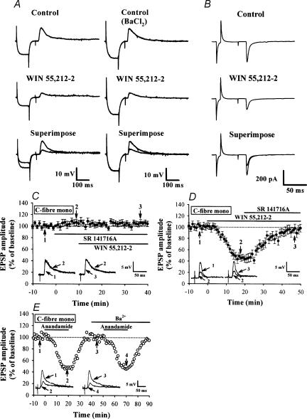
Figure 2. Cannabinoid receptor agonists reduce monosynaptic EPSPs and EPSCs evoked by mandibular nerve stimulation.
A, sample traces showing synaptically evoked EPSPs from cells recorded before and 20 min after application of WIN (5 μm) in the absence (left) or presence of Ba2+ (100 μm) (right). The EPSP was preceded by a transient hyperpolarizing current pulse (0.1 nA, 100 ms) passed through the recording microelectrode to measure input resistance (IR) of postsynaptic neurones. Note that bath application of WIN decreased the amplitude of EPSPs, which was accompanied by a substantial decrease in membrane IR. In the presence of Ba2+, WIN was still able to depress EPSP amplitude without affecting membrane IR. The average resting membrane potential (RMP) and IR of 12 neurones tested were −66.9 ± 2.7 mV and 105.4 ± 3.5 MΩ, respectively, before WIN application, and −68.9 ± 2.5 mV and 99.6 ± 3.2 MΩ after WIN application. B, sample traces showing the effect of WIN (5 μm) on monosynaptic EPSCs. The neurone was voltage clamped at a holding potential of −70 mV, and EPSCs were evoked at 30 s intervals in the presence of strychnine sulphate (0.5 μm), bicuculline methiodide (10 μm) and d-APV (50 μm). Traces are averaged from 6 responses. C, summary time plot of C-fibre EPSPs (n= 5) showing a potentiating effect of SR 141716A (5 μm) on the EPSP amplitude and its blocking effect on the WIN-induced inhibition of EPSPs. D, summary time plot of C-fibre EPSPs (n= 4) showing that SR 141716A (5 μm) reversed the WIN-induced inhibition of EPSPs. E, effect of external Ba2+ on the anandamide-induced inhibition of EPSPs. Ba2+ (100 μm) had no effect on the inhibitory effect of anandamide (30 μm) on monosynaptic C-fibre EPSPs. Superimposed EPSPs were taken at the times indicated. Similar results were also observed in other three cells. Bars indicate the period of drug application.

