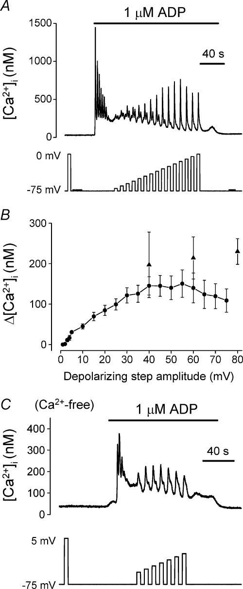
Figure 1. Effect of pulse amplitude on the depolarization-evoked [Ca2+]i increase during stimulation of P2Y receptors.
[Ca2+]i responses of rat megakaryocytes to ADP (1 μm, horizontal bar) and step depolarizations from a holding potential of –75 mV. The effect of depolarization during ADP application was assessed after the agonist-evoked increase had settled to a raised plateau level. A, effect of increasing the amplitude of the depolarizing step in 5 mV increments up to 75 mV. B, relationship between average peak [Ca2+]i response and depolarizing pulse amplitude (mean ±s.e.m.). Data plotted as circles were from two series of increasing amplitude voltage steps; up to 75 mV in 5 mV increments (as shown in A; 18 cells) and up to 5 mV in 1 mV increments (9 cells). Data plotted as triangles represent the average (5–9 cells) maximal [Ca2+]i increase during application of repeated 5 s duration pulses, of either 40, 60 or 80 mV amplitude, at the same frequency as in A. C, example of an experiment demonstrating that depolarization still potentiated ADP-evoked responses in a graded manner in Ca2+-free saline.

