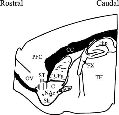
Figure 1. A drawing of a parasagittal slice of the rat brain showing the placement of the stimulating electrode (ST) and the region in which the recordings were performed (hatched area).
Abbreviations: Nac, nucleus accumbens (Sh, shell; C, core); ACP, anterior commissure, posterior; CC, corpus collosum; Cpu, caudate-putamen; Fx, fornix; OV, olfactory ventricle; TH, thalamus; PFC, prefrontal cortex; Hip, hippocampus. (Adapted from Paxinos & Watson, 1998.)

