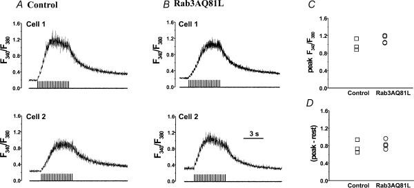
Figure 7. Expression of Rab3AQ81L does not alter global [Ca2+]i dynamics during repetitive stimulation.
A, perforated patch [Ca2+]i recordings from two control cells infected with GFP–IRES–β-galactosidase adenovirus, stimulated with a train of 40 ms depolarizations (20 pulses, −90 to +20 mV, 200 ms interval between pulses). B, [Ca2+]i recordings from two cells infected with Rab3AQ81L–IRES–GFP adenovirus. C, maximal background-corrected fluorescence ratio (‘Peak’) during the train for control (n= 3) and Rab3AQ81L-expressing cells (n= 4). D, maximal change in fluorescence ratio, determined from the difference between the peak fluorescence ratio and the ratio at rest, prior to stimulation. Cells were loaded by preincubation with 2 μm l−1 Fura-4F-AM.

