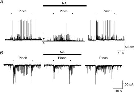
Figure 2. Reduction of pinch-evoked firing of SG neurons by NA.
A, pinch stimuli applied to the ipsilateral hindlimb produced a barrage of EPSPs accompanied by action potentials under a current-clamp condition (left). NA (50 μm) hyperpolarized SG neurone (arrows) and inhibited the action potentials in a reversible manner (middle and left). B, left, EPSCs elicited by pinching in voltage-clamp mode (VH=−70 mV). NA (50 μm) suppressed repeated EPSCs during pinching in a reversible manner without affecting amplitude of large EPSCs evoked at the beginning and end of pinch stimulus (middle and right).

