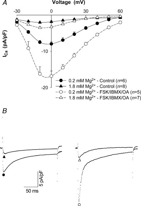
Figure 4. Effect of [Mg2+]p on ICa in high phosphorylation conditions.
A, current–voltage relationships for ICa in myocytes dialysed with 0.2 mm and 1.8 mm[Mg2+]p in control (continuous curves) and high phosphorylation conditions (dashed curves) in the absence and presence of 10 μm forskolin (FSK), 300 μm IBMX and 50 μm OA, respectively, when [Ca2+]o was set at 0.5 mm. Currents were recorded at 5 min after break-through into the whole-cell patch-clamp configuration. Data are means and s.e.m., with the number of experiments indicated in parentheses. B, tracings of typical ICa records at a test potential of 0 mV in rat ventricular myocytes dialysed with 0.2 mm (•) and 1.8 mm[Mg2+]p (▴) in control conditions, and 0.2 mm (○) and 1.8 mm[Mg2+]p (▵) in high phosphorylation conditions.

