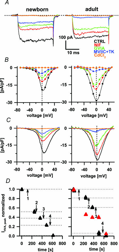
Figure 2. The effects of nifedipine, ω-conotoxin GVIA, ω-conotoxin MVIIC with ω-agatoxin TK and CdCl2 on Ba2+ currents.
In panels A, B, C and D left plots present data from the newborn and right plots from the adult animals. A, Ba2+ currents were evoked during 30 ms voltage steps from −80 mV to +60 mV. All recordings were obtained after preincubation (2 min) with different VACC blockers as indicated. B, current densities from the same cells were plotted to obtain the Ba2+ current I–V relationship. C, the effect of VACC blockers on the Ba2+ current I–V relationship obtained from 300 ms voltage ramps. D, the time course of the effect of nifedipine (1), GVIA (2), MVIIC/TK (3) and CdCl2 (4) on Ba2+ currents. LVA currents in adults are shown as red triangles and the HVA are shown as black triangles. Note that current values were normalized to the peak amplitude of both VACC components. Nifedipine blocked about 50% of the LVA component; however, other toxins than Cd2+ did not affect the LVA component.

