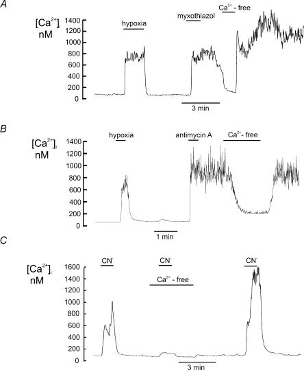
Figure 3. Effects of electron transport inhibitors on [Ca2+]i in type I cells.
Effects of electron transport inhibitors on intracellular calcium in type I cells (measured using indo-1) in the presence and absence of extracellular calcium. A, myxothiazol (1μm) caused a large increase in intracellular calcium which was slow in onset, often with a delay of 30 s to 2 min, and which was maintained even after wash out of the drug. This sustained increase in [Ca2+]i was substantially reduced by removal of extracellular Ca2+. B, antimycin A (0.5μm) caused a rapid increase in [Ca2+]i that was also maintained following wash out of the drug. The antimycin A induced increase in [Ca2+]i was largely reversed by removal of extracellular Ca2. C, cyanide (nominally 2 mm; see methods) caused a rapid and reversible increase [Ca2+]i. The cyanide induced increase in [Ca2+]i was greatly reduced in calcium free media.

