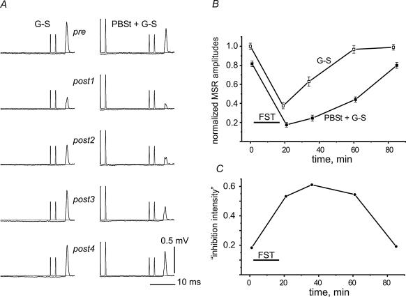
Figure 3. Postfatigue changes in the presynaptic inhibition of G–S MSRs induced by conditioning stimulation of the nerve to the posterior biceps and semitendinosus muscle (PBSt).
A, fatigue-evoked changes in G–S MSRs (averages of 12 records in each test interval) without (left column) and with (right column) preceding stimulation of the PBSt nerve. The conditioned reflexes were obtained around 30 s after the control reflexes. The intensity of the PBSt stimulation was set to evoke approximately 20% inhibition of the G–S MSR in the prefatigue tests. B, amplitudes of the unconditioned (G–S) and conditioned (PBSt + G–S) reflexes in different pre- and postfatigue test periods, normalized to the mean value of the prefatigue G–S MSR. C, ‘Inhibition intensity’, as defined in the text, plotted against time.

