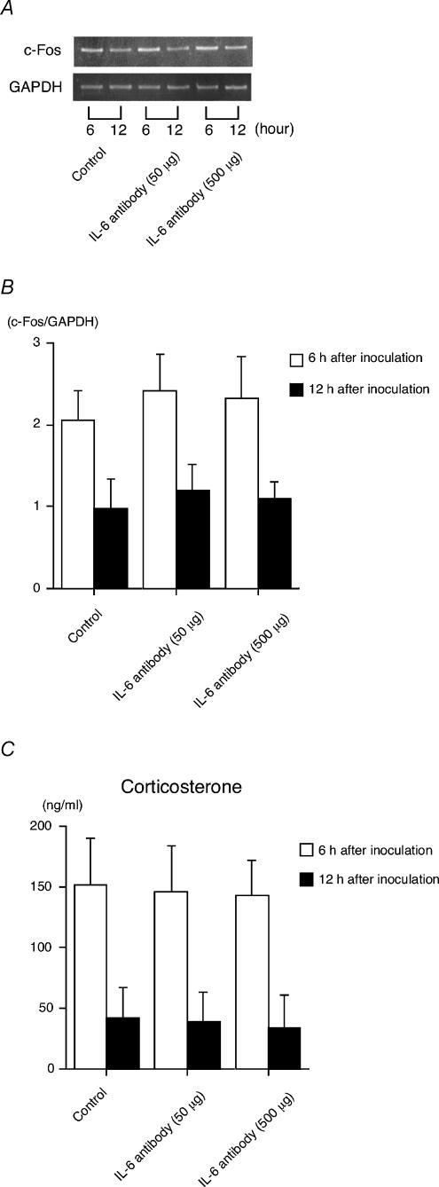
Figure 5. Effects of anti-IL-6 treatment on the c-Fos expression in the paraventricular nucleus and the corticosterone response in the plasma.
GF mice at 5 weeks of age were injected intraperitoneally with either anti-IL-6 antibody (MP5-20F3; 50 or 500 μg) or control rat IgG antibody (control) 1 h before being inoculated with Bifidobacterium infantis. The analysis of c-Fos mRNA expression levels in the paraventricular nucleus was done at 6 or 12 h after the inoculation. A, the results shown are representative of 4 independent experiments. B, histogram shows the relative band intensities on densitometric analysis as ratios of c-Fos and GAPDH mRNA after 30 cycles of amplification (n = 4 for each time point). C, determination of the plasma corticosterone levels was carried out according to the protocol described in the Methods (n = 6 for each time point).

