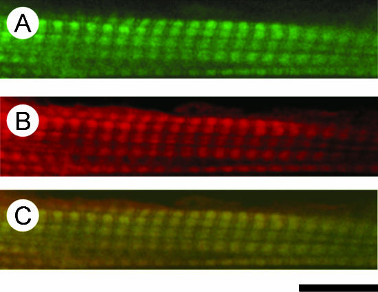
Figure 2. Confocal fluorescence micrographs of bovine myocardium reconstituted with Rh-IA-labelled Tm and labelled with Fl-Ph.
The thin filament was first removed by gelsolin, and sequentially reconstituted with actin, followed by Rh-IA-labelled Tm. The preparation was then treated with Fl-conjugated phalloidin to label the actin filament. A, Fl fluorescence showing the distribution of the actin filament; B, Rh fluorescence showing the distribution of Tm; C, colocalization of the actin filament and Tm. Calibration bar, 10 μm.

