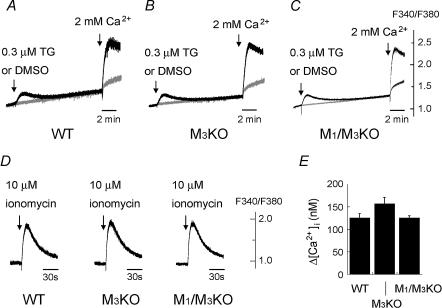
Figure 4. Effect of thapsigargin (TG) and induction of capacitative Ca2+ entry (A–C), and Ca2+ release from the internal Ca2+ store by ionomycin (D and E) in WT, M3KO and M1/M3 double KO SMG cells.
Drugs were applied to the SMG cell suspension at the time point indicated by the arrow. A–C, [Ca2+]i change in the nominal absence of external Ca2+ in response to 0.3 μm thapsigargin (black trace) or control dimethylsulfoxide (DMSO; grey trace) and that caused by addition of 2 mm Ca2+. Note the large [Ca2+]i increase induced in all of the WT (A), M3KO (B) and M1/M3 double KO (C) SMG cells by adding external Ca2+ following TG application (capacitative Ca2+ entry). D, [Ca2+]i change in the absence of external Ca2+ in response to 10 μm ionomycin in the WT, M3KO and M1/M3 double KO SMG cells. 1 mm EGTA was added to the external solution. E, summarized peak [Ca2+]i increases induced by 10 μm ionomycin in WT, M3KO, or M1/M3 double KO SMG cells (mean ± s.e.m.; n = 4 for each genotype). All the results shown are representative of 4 experiments.

