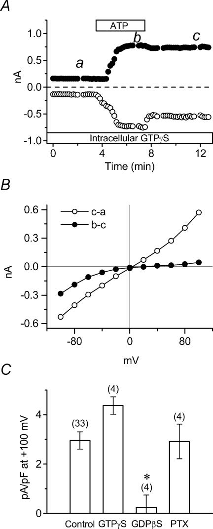
Figure 3. Role of G-protein coupled purinergic receptors in activation of ICl,ATP.
A, time course of extracellular ATP-induced whole-cell currents in standard extracellular solution during application of 100 μm ATP to the bath. The tested cell was dialysed with 0.1 mm GTPγS, and was exposed to ATP as indicated by the bar. B, I–V relationships of ATP-induced currents (b–a) and the persistently activated ICl,ATP (c–a). Whole-cell currents were activated by voltage-clamp pulses as in Fig. 1B, at time points a, b and c in A. C, mean current densities at +100 mV of ICl,ATP in GTPγS-dialysed (n = 4), GDPβS-dialysed (n = 4) or PTX-pretreated (n = 4) cells. * signifies significantly smaller than control with P < 0.05.

