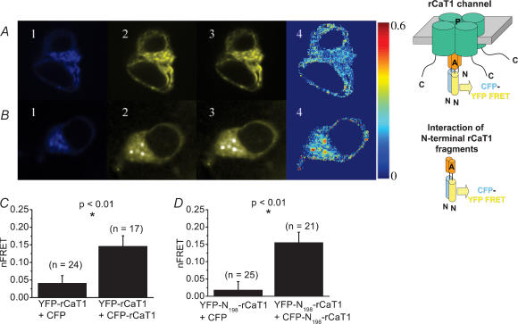
Figure 4. In vivo assembly of CFP-/YFP-labelled rCaT1 proteins and of their CFP-/YFP-labelled N-terminal fragments.
HEK cells were cotransfected with CFP- and YFP-labelled CaT1 (A) or CFP- and YFP-labelled N-terminal rCaT1 fragments (B). Representative CFPexcit/CFPemis (1), CFPexcit/YFPemis (2, FRET), and YFPexcit/YFPemis (3) images and the corrected FRET image (4, nFRET) are shown in A and B. For control, transfection was carried out with CFP and YFP-rCaT1 (C) or CFP and YFP-N198-rCaT1 (D). On average, CFP-/YFP-rCaT1 (C) and CFP-/YFP-N198-rCaT1 (D) cotransfected cells showed a significantly higher nFRET when calculated for the whole HEK cell in comparison to control. n denotes the number of cells analysed.

