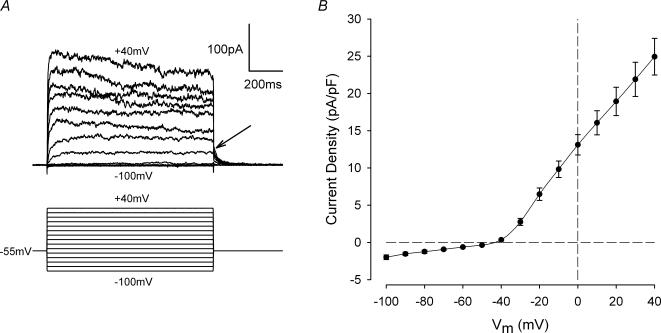
Figure 1. Current—voltage relation of acutely isolated chondrocytes.
A, representative family of membrane currents from a voltage-clamped articular chondrocyte. The voltage-clamp protocol is shown below the current records. Holding potential (Vh) was −55 mV, and the membrane potential was stepped from +40 to −100 mV in 10 mV increments at 3 s intervals. Arrow indicates deactivation of outward currents at the end of the depolarizing steps. Cell capacitance was 6.5 pF. B, mean peak current—voltage relationship from 24 cells (5 different cell preparations). Currents from each cell were normalized to cell capacitance, and then averaged. The mean cell capacitance of the 24 cells was 5.8 ± 0.3 pF.

